
Reviewed by Dr Edgar Tay, interventional cardiologist and structural heart specialist at Asian Heart & Vascular Centre.
Last reviewed September 2026.
Reading time: about 11 minutes.
TL;DR
A clot in a deep leg vein (deep vein thrombosis, or DVT) and a clot in the lung circulation (pulmonary embolism, or PE) are two presentations of one condition, venous thromboembolism. Confirming one follows a fixed sequence: a structured probability assessment, a D-dimer blood test that is most useful when negative, then leg ultrasound or a CT scan of the lung arteries. Singapore’s national guidance is to start anticoagulation as soon as a proximal DVT or PE is confirmed, and to continue it for at least three months. Most patients are treated at home. Assessment at AHVC in Singapore establishes which path applies to you.
The situation you are probably in
Most people arrive through one of four doors.
A calf that has been swollen and aching for three or four days, put down to a training session or a long drive, and has not settled. One leg only, larger than the other, warm, tender along the inner calf.
Breathlessness that arrived without warning, sometimes after a long flight or a stretch in hospital, with a sharp catch at the top of each breath.
A scan ordered for something else, reporting a clot in a lung artery nobody was looking for.
Or a telephone call from a GP the morning after a blood test, asking you to come in that day.
They feel completely different, and they are the same question: is there a clot, and what happens next. This article answers the second half. If what you want first is how a clot forms, that is in our guide to thrombosis.
Two clots, one disease, and why that changes the plan
A clot in a deep leg vein is a DVT. A clot that has travelled to the lung arteries is a PE. They are investigated and treated as one condition, venous thromboembolism, because in most patients they are the same event at two points in time.
When patients admitted with a symptomatic PE have both legs examined with ultrasound, a DVT is found in 70.6 per cent of them, and in 57.3 per cent of those cases the leg was producing no symptoms at all. The clot in the lung had travelled from a leg that felt normal.
Two things follow. A leg clot is not a local problem, because it has an open route to the lungs, which is why a newly swollen calf is investigated rather than watched for a week. And a lung clot does not need leg symptoms to have started in the leg, so a normal-looking leg does not argue against the diagnosis.
What a PE does inside the lung, and how it differs from pulmonary hypertension, are covered on the pages for pulmonary embolism and the difference between pulmonary embolism and pulmonary hypertension.
When is a blood clot an emergency, and where should you go?
Some presentations are assessed the same day, in an emergency department rather than an outpatient clinic:
- Breathlessness that came on suddenly, or that is worsening hour by hour.
- Sharp chest pain that is worse on breathing in.
- Coughing up blood.
- Fainting, or feeling close to fainting, with any of the above.
- One leg that has become swollen and painful over hours to days, particularly with redness or warmth.
In Singapore, that means calling 995 or going to the nearest emergency department. Asian Heart & Vascular Centre is a specialist outpatient practice and is not the route for an unstable patient.
One misconception is worth correcting here. A PE is often expected to feel like a heart attack, with crushing central chest pain. It frequently does not. The common picture is breathlessness out of proportion to what you were doing, with a catch at the end of a deep breath. Waiting for chest pain that never arrives is how the diagnosis gets delayed.
How a clot is confirmed
Step one: structured clinical probability. Before any test, the assessing doctor scores the likelihood using a published rule such as the Wells or revised Geneva score. Among patients scored low, about 10 per cent turn out to have a PE, rising to around 30 per cent in the moderate category and 65 per cent in the high category. The score decides which test comes next and how to read it.
Step two: D-dimer, which is most useful when it is negative. D-dimer measures a fragment released when the body breaks down a clot, and modern assays have a diagnostic sensitivity of 95 per cent or better. A normal result in a patient already assessed as low or intermediate probability is strong evidence against a clot: the risk of a thromboembolic event in that group over the next three months without treatment is under 1 per cent. Over the age of 50, an age-adjusted threshold of age multiplied by 10 micrograms per litre reduces unnecessary scanning.
A raised D-dimer is not a diagnosis. It rises in infection, after surgery, in pregnancy, in cancer and with age. Reading it as a clot test rather than an exclusion test is the commonest misreading of this pathway.
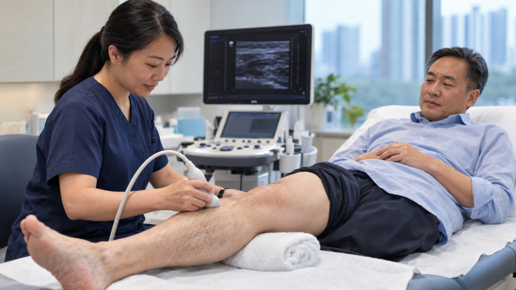
Step three: imaging the leg, or the lung. For a suspected leg clot, the test is compression ultrasound, with a sensitivity and specificity above 95 per cent for a proximal DVT. The sonographer presses a probe along the vein at intervals: a healthy vein flattens; one containing a clot does not. No needles, no radiation, no preparation. For a suspected lung clot, the test is CT pulmonary angiography, the method of choice for imaging the lung circulation, with a sensitivity of 83 per cent and a specificity of 96 per cent in the PIOPED II study. It involves a cannula, iodine-based contrast, and a breath-hold.
An echocardiogram is often added once a PE is confirmed. It does not make the diagnosis. It shows whether the right side of the heart is under strain, which separates a patient who can go home from one who cannot.
Where this happens in Singapore
Venous thromboembolism is less common in Asian populations than in Western ones, and it is not rare here. European figures put the annual incidence of PE at 39 to 115 per 100,000 people and of DVT at 53 to 162 per 100,000. The published population-based estimate for Singapore, from National Healthcare Group data for 2006, is 57 per 100,000 for venous thromboembolism and 15 per 100,000 for PE. That figure is dated, and it remains the local reference point.
Patients reach the diagnosis by three routes here: through a GP or polyclinic that orders a D-dimer or refers for an ultrasound, through an emergency department, where most confirmed PE begins, or as an incidental finding on a scan done for cancer staging or post-operative follow-up.
A same-week leg ultrasound depends on three things: a referral, a slot in a vascular laboratory or radiology department, and a clinician free to act on the result the day it returns.
Both the public and private systems carry this pathway. The Agency for Care Effectiveness, the health technology assessment body of the Ministry of Health, publishes national guidance on anticoagulant choice and duration. Its position is to start anticoagulation as soon as possible once a proximal DVT or PE is confirmed, unless there is a contraindication.
Treatment, matched to the clot
Anticoagulation is the backbone, and for most patients it is the whole of the treatment. Singapore guidance is to use a direct oral anticoagulant (DOAC) for at least three months as the preferred option, with warfarin, a vitamin K antagonist, as the alternative where a DOAC is unsuitable. International guidance agrees. Low molecular weight heparin injections retain defined roles, including in cancer-associated clot and in pregnancy.
Most people are not admitted to hospital. International guidance suggests home treatment over hospital treatment for DVT and for low-risk PE, and European guidance carries a specific recommendation for early discharge in low-risk PE. Admission is reserved for unstable patients, need oxygen, have a high bleeding risk or significant kidney impairment, or whose circumstances make outpatient monitoring unsafe. A confirmed clot no longer means an automatic hospital stay.
At the severe end, treatment is layered rather than substituted. For a PE causing circulatory compromise, thrombolytic therapy followed by anticoagulation is recommended over anticoagulation alone. Where the heart shows strain but blood pressure is maintained, anticoagulation alone is suggested over routine thrombolysis, because the bleeding cost outweighs the benefit. Where thrombolysis is contraindicated or has failed, surgical embolectomy and percutaneous catheter-directed treatment are the options, described on the page for pulmonary thrombectomy.
For a leg clot, intervention is the exception. Guidance suggests anticoagulation alone over thrombolytic therapy for proximal DVT. The ATTRACT trial tested that directly: adding pharmacomechanical catheter-directed thrombolysis produced post-thrombotic syndrome in 47 per cent of patients against 48 per cent on anticoagulation alone, with major bleeding in 1.7 per cent against 0.3 per cent. Clot removal is reserved for a defined minority, typically extensive iliofemoral clot with a threatened limb.
Breathlessness persisting months after a PE is a separate question. Assessment for chronic thromboembolic pulmonary hypertension is recommended where symptoms continue or return. That is the chronic end of this story, with its own procedures, including balloon pulmonary angioplasty.
How long will you be on blood thinners, and what decides it?
Every patient with a confirmed proximal DVT or PE is anticoagulated for a minimum of three months, to prevent the clot extending and to prevent a recurrence. International guidance suggests a shorter primary course of three to six months over a longer one of six to twelve months. What matters is not the length of that first course but what happens at the end of it, and that turns on one question: why did this clot form?
Provoked by a major transient risk factor, such as surgery, immobilisation, significant trauma or a plaster cast. The provoking factor has gone, and anticoagulation usually stops at the end of the primary course.
Provoked by a chronic risk factor, or unprovoked. Where the risk factor persists, or where no cause is found at all, guidance suggests indefinite antithrombotic therapy over stopping. Singapore guidance frames it the same way: unprovoked venous thromboembolism carries a higher recurrence risk and may require extended treatment where bleeding risk is low or moderate.
The numbers are worth seeing. After stopping anticoagulation following a first unprovoked clot, recurrence runs at 10.3 events per 100 person-years in the first year, and cumulative incidence reaches 16 per cent at two years, 25 per cent at five years and 36 per cent at ten years. Of those recurrences, 4 per cent are fatal. Against that, major bleeding occurs in roughly 1 to 3 per cent of patients per year on a vitamin K antagonist, and about 30 per cent less often on a DOAC.
“Indefinite” does not mean the decision is never revisited. The default becomes continuation, reviewed as bleeding risk, kidney function, age and other medicines change.
What happens afterwards
Post-thrombotic syndrome is the common long-term consequence, and it is under-discussed. Aching, heaviness, swelling and skin changes in the affected leg develop in 20 to 50 per cent of patients after a proximal DVT, and the syndrome can only be diagnosed three to six months after the event, once the acute symptoms have settled.
Compression stockings do not reliably prevent post-thrombotic syndrome, although patients are often told they will. Current guidance suggests against their routine use to reduce that risk, while noting that stockings may still reduce swelling and pain in selected patients during the acute phase. They are a symptom measure rather than a preventive one, worth wearing if they make the leg feel better.
Exercise is not restricted by the clot. Walking is encouraged once treatment has started. The constraint during anticoagulation is bleeding, which is why contact sports and high-fall-risk activities are worth raising at the consultation. Flying depends on how long ago the clot was, whether you are still anticoagulated, and what provoked it, and the travel evidence is covered in our article on blood clots and long flights.
Thrombophilia testing is usually not the answer. Patients with an unexplained clot often ask to be screened for an inherited clotting disorder, and whether their children should be. Current guidance suggests not performing thrombophilia testing to guide the duration of anticoagulant treatment in unprovoked venous thromboembolism, and likewise in surgically provoked venous thromboembolism. The result rarely changes what you are advised to do.
One connection to a different pathway is worth naming once. Where a persistent opening exists between the upper chambers of the heart, a clot from the leg veins can cross to the arterial side and reach the brain rather than the lungs. That route is set out in our guide to a hole in the heart (PFO). A clot that forms inside the heart itself arrives by a different route, described on the cardioembolic stroke page.
Realistic expectations, and what anticoagulation does not do
Anticoagulation does not dissolve the clot you already have. This is the commonest misunderstanding of the treatment, and the name “blood thinner” causes it. What anticoagulation does is prevent the clot from extending and prevent a new one from forming. The existing clot is broken down by the body’s own fibrinolytic system over weeks to months, often incompletely, so an ultrasound a year later may still show an abnormal vein in a patient who is entirely well.
The trade-off is bleeding. Easier bruising, longer bleeding from small cuts, and heavier menstrual bleeding are expected. Anyone on extended treatment should know what to do about a significant bleed and who to contact.
Good treatment does not remove the long-term risk. Post-thrombotic syndrome develops despite correct anticoagulation, adding catheter-directed thrombolysis does not prevent it, and breathlessness on exertion can persist for months after a PE.
What a consultation involves
Bring the material the decision is built on: the ultrasound or CT report, the discharge summary if you were admitted, the name and dose of the anticoagulant and the date you started it, and your other medicines including hormonal contraception.
You should leave knowing four things: whether your clot is provoked or unprovoked and why, which anticoagulant you are on and for how long, the date of the review that decides whether treatment continues, and what symptoms should bring you back sooner.
Useful questions to ask:
- Was this clot provoked, and by what?
- What happens at the three-month review, and who makes that call?
- What is my bleeding risk on this medication?
- When can I fly, exercise, or return to normal work?
Who does this at Asian Heart & Vascular Centre
Asian Heart & Vascular Centre is a specialist cardiology practice with 10 cardiologists working across five hospital locations in Singapore. Dr Edgar Tay, interventional cardiologist and structural heart specialist, works in structural and pulmonary vascular intervention, and his practice includes balloon pulmonary angioplasty for chronic thromboembolic pulmonary hypertension. Echocardiography for right heart strain sits within the clinic’s investigations.
Where the answer is somewhere else, it is worth saying so.
- An unstable patient belongs in an emergency department, and that admission is managed by the hospital team.
- Leg vein ultrasound and CT pulmonary angiography are arranged through a hospital radiology department or vascular laboratory.
- Anticoagulation in pregnancy, in cancer-associated thrombosis, or where an inherited clotting disorder is suspected is typically co-managed with haematology.
- Extensive iliofemoral clot considered for removal involves vascular surgery or interventional radiology.
If you have been told you have a clot in your leg or your lung, the questions that matter next are why it happened, how long you will be treated, and who reviews that decision. A consultation with Dr Edgar Tay at Asian Heart & Vascular Centre can work through those with you, and say plainly where another specialty should take the lead.